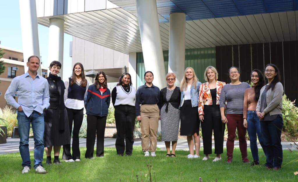
Professor Caroline Gargett has spent decades working to address the two major issues presented by endometriosis: diagnostic delay and poor treatment options.
Endometriosis is a disease affecting women where tissue similar to the lining of the womb grows outside the womb, in other parts of the body. Symptoms include severe and chronic pelvic pain, and while endometriosis most often affects the reproductive organs, it is frequently also found in the bowel and bladder and has occasionally even been found in muscles, joints, the lungs and the brain.
Endometriosis is a “quality of life” disease and numerous international studies have demonstrated that the long diagnostic delay impacts women’s health and wellbeing, including social activities, mental and emotional health, education, employment, finances and sexual relationships.
Prof Gargett is a world leader in this area – her international standing was recognised by the Endometriosis Foundation of America, which named her among the inaugural members of its Scientific Advisory Board. She also has a position on the International Scientific Committee of the Fondation pour la Recherche sur l’Endométriose in France.
In 2019, Professor Caroline Gargett and collaborators at the University of Queensland and Monash IVF were the first to be awarded funding from the United States Department of Defense for endometriosis research (AU$3.05 million over 4 years) to determine the cause and the physiological processes associated with the disease.
In a study published in Reproductive Biomedicine Online, Prof Gargett’s team was the first in the world to show the role of endometrial stem/progenitor cells in the disease – establishing that they can escape in menstrual fluid from the uterus through the fallopian tubes into the pelvic cavity, where they have the potential to survive and grow into painful lesions.
Using menstrual fluid to diagnosis endometriosis
Prof Gargett is currently working on a non-invasive diagnostic technique, using a woman’s own menstrual fluid to determine the presence of endometriosis. At present, apart from imaging, which cannot identify all types of endometriosis, surgery is still the only method of establishing a definitive diagnosis and the only way to remove the lesions which cause so much pain and distress. However, they frequently return in most patients.
Australian Menstrual Fluid Biobank for Endometriosis Research
Key to the success of this project is establishment of the Australian Menstrual Fluid Biobank for Endometriosis Research (AMBER), a sustainable, living biobank of five menstrual fluid components per sample, to enable the development of new treatments and diagnostics for endometriosis and potentially other gynaecological conditions.

Prof Gargett’s aim is to dramatically reduce the time to diagnosis from the current 6.5 years and for pathology services to analyse menstrual fluid samples as commonly as blood tests.
“Menstrual fluid contains endometrial tissue and provides a non-invasive way of obtaining this tissue,” Prof Gargett said. “We want to develop a diagnostic test for endometriosis based on its cellular, protein or molecular components.”
An added bonus of the Biobank concept is that these samples could also be used to generate 3D endometrial organoids and stromal cultures, which can then be tested to identify the most suitable treatment for each individual patient.
Prof Gargett and her team of researchers also intend to develop a non-invasive alternative for menstrual fluid collection.
This ambitious project could one day be recognised as the crucial point at which both diagnosis and treatment of endometriosis became quicker, easier and more personalised.


